

Researchers on the College of Osaka have developed a brand new know-how that permits the precise visualization of various elements that make up a single cell
The tissues encompass a heterogeneous combination of several types of cells, complicating our understanding of their organic capabilities and research on the illness. Now, a multi-institutional workforce led by the College of Osaka has developed and supplied the proof of a brand new know-how to view the distribution of parts in a single cell, opening the best way of a a lot larger understanding of ailments in advanced organic samples.
T-Spsi (ionization of tapping mode scanning) is a way that permits the evaluation of the molecules in a pattern. A number of micro-samples of the totally different areas of a cell are taken and transferred for evaluation by a way referred to as mass spectrometry, which may decide the precise chemical parts of that area. “We’ve developed a brand new T-Spes unit that permits us to visualise the microscopy pattern in a number of methods,” explains the principle writer Yoichi Osuka. “We are able to additionally observe the sampling course of instantly as micro-samples are taken to research mass spectrometry.”
By modifying the beforehand developed T-Spesi know-how, the workforce allowed the analytical unit to be positioned above a reversed fluorescence microscope. This enables to look at the sampling course of, in addition to the direct remark of the pattern itself. The pattern will be imagined in numerous methods, permitting the detection of any fluorescent -labeled goal molecules, figuring out the distribution of traits on the cell floor and the picture of the areas of the cell chemical parts.
This know-how can visualize the distribution of intracellular lipids, the fats compounds that play key roles in metabolic processes. It’s recognized that irregular distributions and capabilities of lipids are associated to the illness. “Once we utilized our know-how to mannequin the cells, we might see the lipids from every particular person cell utilizing the imaging imaging, instantly view the cell by fluorescent microscopy and likewise decide the floor type of the cell,” explains the senior writer Michisato Toyoda. They have been additionally capable of detect distinctions between several types of cells with totally different mobile compositions. “This enables an understanding of the multidimensional molecular info of the person cells inside a sick tissue pattern,” says Osuka.
This new attention-grabbing know-how could have an awesome affect on our capacity to grasp the processes underlying the illness improvement in advanced organic samples, permitting us to grasp the advanced mixtures and interactions of the cells current within the tissue samples. This may contribute to the event of superior therapies and diagnostic strategies for all kinds of ailments.

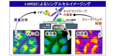
Fig. 1
The schematic diagram of a single cell MSI utilizing the ionization of the scanning electrospray in tapping mode.
Fig. 2
Block diagram of newly developed measurement system and a picture of the machine measuring Hela cells
Determine three
A graph that reveals the outcomes of the evaluation of two sorts of hela cells
The article, “Imagistics of mass spectrometry with a single lipid cell cell in Hela cells via ionizing the tapping mode scan”, was printed within the chemistry of communications: https://doi.org/10.1038/S42004-025-01521-2
/Public launch. This materials from the Authentic Authentic Group (s) (the writer) could possibly be punctual and edited for readability, type and size. Mirage.Information doesn’t take institutional positions or sides and all of the opinions, positions and conclusions listed here are solely these of the writer (writer).